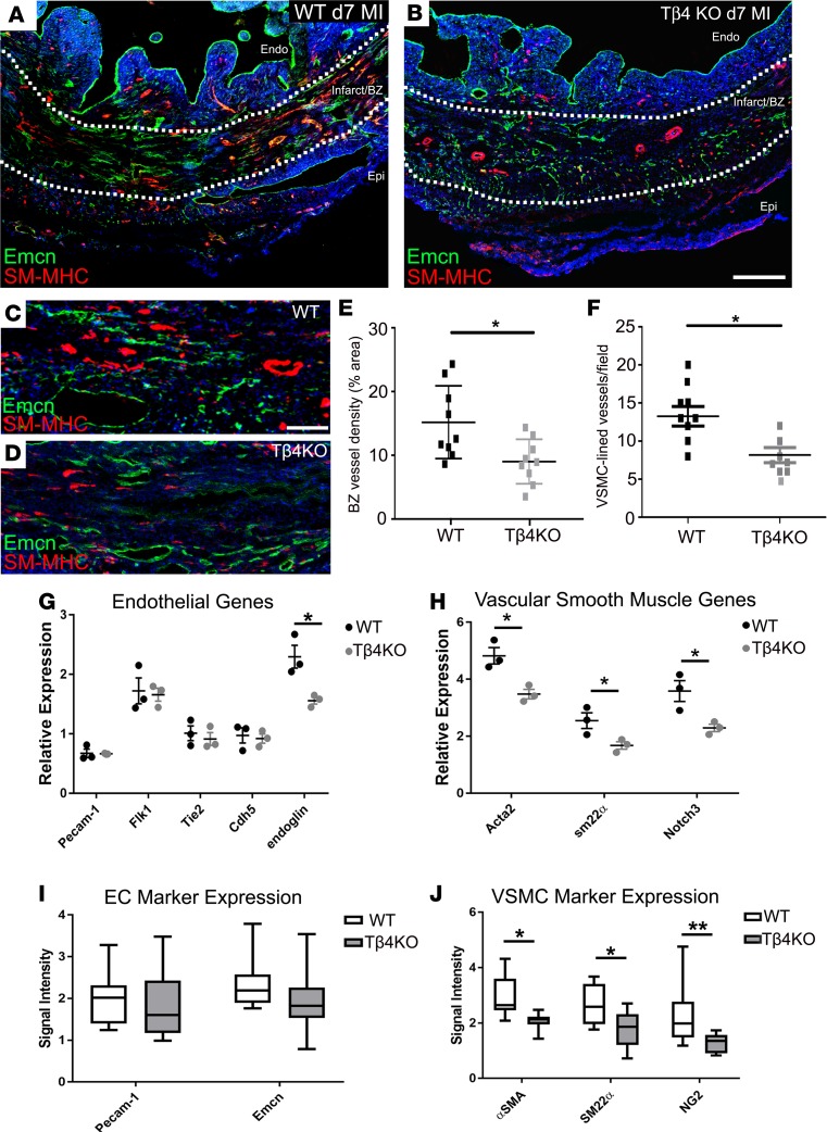
Figure 12. Neovascularization is diminished in the infarct border zone of thymosin β4 KO hearts.
By immunofluorescence, vascular density within the border zone was significantly reduced in Tβ4KO hearts, compared with WT (A–D, quantified in E), and vessels appeared less mature (B, compared with A). Fewer vessels acquired smooth muscle support (D, compared with C, quantified in F). By qRT-PCR, endothelial cell markers were not significantly reduced, with the exception of endoglin (G), whereas smooth muscle markers, Acta2, Sm22a and Notch3, were all significantly reduced in Tβ4KO hearts (H). These differences were reflected in quantification of immunofluorescence signal intensity, showing no significant reduction of endothelial markers (I) but a significant reduction of smooth muscle markers (J). Sections are representative of n = 9 hearts per genotype and n = 9 quantified. BZ, border zone; endo, endocardium; epi, epicardium. Scale bars: 500 μm (A and B); 50 μm (C and D). Statistical analyses (E and F): Mann-Whitney test (2-tailed); (G–J) 2-way ANOVA with Bonferroni correction for multiple comparisons; scatter plots: each data point represents a separate animal with mean ± SEM; box-and-whisker plots show mean ± minimum/maximum; *P ≤ 0.05, **P ≤ 0.01.

