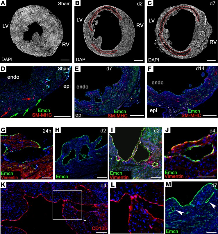
Figure 3. Remodeling of the endocardium after myocardial infarction.
Immunostaining revealed increased trabeculation of the endocardial surface following MI: a representative sham-operated heart (A), compared with MI hearts after 2 (B) and 7 (C) days. Whereas, in the uninfarcted heart, large coronary arteries (red arrows) and veins (green arrows) were restricted to the epicardial side of the ventricle (D), new vessels appeared on the endocardial side of the infarct (E and F), coincident with compaction of trabeculae. Altered endocardial morphology was detected as early as 24 hours, with formation of cavities and finger-like protrusions (G–I). Coalescence of endocardial cells and trabecular compaction between days 4 and 7 (J–M; box in K enlarged in L) coincided with appearance of subendocardial vessels (arrowheads, M). LV, left ventricle; RV, right ventricle; epi, epicardium; endo, endocardium. Scale bars: 1 mm (A–F); 100 μm (G, K, and J); 200 μm (H, I, and M). The boxed area in K is magnified 2-fold in L. Representative of 24 hours: n = 5 (2 sham); 2 days: n = 6 (3 sham); 4 days: n = 5 (2 sham); 7 days: n = 15 (4 sham); 14 days: n = 5 (2 sham).

