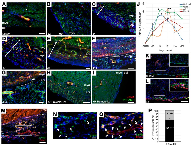
Figure 7. Growth of a vascular network after myocardial infarction within the reactivated epicardium.
Shown by immunostaining, the single-cell layer epicardium of the noninjured mouse heart (A) expands rapidly in response to injury, from day 2 after MI (B), continuing to day 14 (C–F). A network of capillaries arises within the activated epicardium between day 4 and day 7 (PECAM1+ endothelial cells in green; αSMA+ vascular smooth muscle cells in red (C–E). Epicardial capillaries remodel to form arterioles (F and G), acquiring smooth muscle support (D–H) and connecting with the underlying coronary vasculature (arrowhead in G). Maximal epicardial activity was observed in the proximity of the infarct (H), with only modest thickening over remote regions of the LV (I). Representative of 24 hours: n = 5 (2 sham); 2 days: n = 6 (3 sham); 4 days: n = 5 (2 sham); 7 days: n = 15 (4 sham); 14 days: n = 5 (2 sham). qRT-PCR confirmed the reexpression of fetal epicardial genes, Wt1, Tbx18, Tcf21, and Aldh1a2 (J, fold change relative to day 2 MI; n = 4 separate animals per time point; mean ± SEM; 2-tailed Kruskal-Wallis nonparametric test with Dunn’s post-hoc test for multiple comparisons; *P ≤ 0.05, ***P ≤ 0.001). WT-1 reactivation in the epicardium, comparing sham (K) and day 2 after MI (L) hearts by immunostaining (boxes in K and L correspond to enlarged insets). PdgfbCreERT2; R26R-EYFP pulse-labeling experiments (M; box enlarged in N and O) indicate that 30.1% ± 3.8% of endothelial cells within the expanded epicardium derived from nonendothelial progenitors or endocardium (arrows in N and O indicate EYFP+ cells; arrowheads indicate EYFP– cells; quantified in P). LV, left ventricle; epi, epicardium; myo, myocardium. Scale bars: 50 μm (A–E, G, and M); 100 μm (H, K, and L); 200 μm (F and I); 20 μm (N and O).

