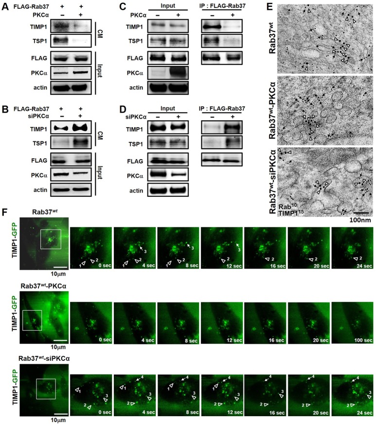
Figure 3. PKCα expression impaired Rab37-mediated exocytotic transport of TIMP1 and TSP1.

(A) PC-14 cells stably expressing FLAG-tagged Rab37 (FLAG-Rab37) were transfected with empty or PKCα-expressing vectors. The cell conditioned medium (CM) was harvested and subjected to immunoblotting. (B) siRNA against PKCα was introduced into cells stably expressing FLAG-Rab37, the TIMP1 and TSP1 levels in CM was analyzed by immunoblotting. (C and D) Cells stably expressing FLAG-Rab37 were transfected with expressing vector (C) or siRNA oligos of PKCα (D). Vesicles were isolated by centrifugations and subjected to IP with anti-FLAG antibody followed by blotting for FLAG-Rab37 and endogenous TIMP1 and TSP1. (E) Cells stably expressing FLAG-Rab37wt were transfected with empty (upper) or PKCα-expressing vector (middle) or siRNA against PKCα (lower). The co-localization of TIMP1 (15 nm of gold, triangle) and Rab37 (10 nm of gold, arrow) in vesicles was observed in immuno-EM images. Scale bars 100 nm. (F) Selected frames from time-lapse movies of TIMP1 trafficking in cells expressing FLAG-Rab37wt, and GFP-TIMP1 together with overexpression (Rab37wt-PKCα) or knockdown (Rab37wt-siPKCα) of PKCα were shown. The numbers labeled horizontal (arrow) or vertical (triangle) movement of GFP-TIMP1. Enlarged images of the boxed areas from time-lapse movies with time intervals in seconds shown. Scale bars 10 µm.
