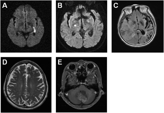
Figure 1. Two GBM cases are diagnosed previously with ischemic stroke using brain MRI images.

The brain MRI images indicate a 72 year-old male patient with the left internal capsule acute infarction on January 21, 2007 (A) and the right hypothalamus acute infarction on April 2, 2009 (B), as well as the right temporal GBM on June 21, 2016 (C). The brain MRI images reveal a 67 year-old female patient with the bilateral frontoparietal subcortical old infarction (D) and the cerebellar vermis GBM on May 21, 2015 (E).
