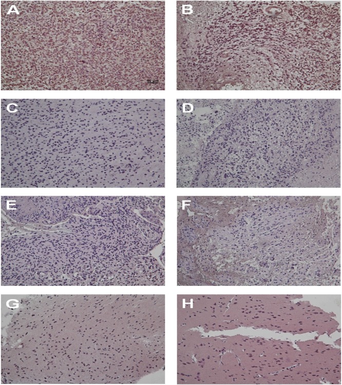
Figure 2. Tumor specimens from GBM patients previously with ischemic stroke display strong HIF-1α expression by IHC staining.

The IHC staining images indicate the expression level of HIF-1α in GBM patients previously with ischemic stroke (A and B), GBM patients previously without stroke (C, D and E) and grade I astrocytoma patients (F, G and H).
