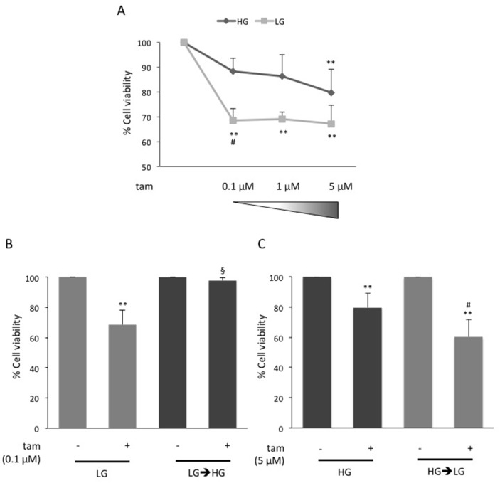
Figure 1. Effect of glucose on MCF7 cell responsiveness to tamoxifen.
(A) MCF7 cells grown in high glucose (25mM; HG) or in low glucose (5.5mM; LG), were treated with estradiol (100nM; E2) and raising concentration (0.1μM, 1μM, 5μM) of tamoxifen (tam); (B) LG cells were shifted to high glucose (LG→HG) during the treatment with E2 and 0.1μM tam; (C) HG cells were shifted in low glucose (HG→LG) when treated with E2and 5μM tam. For all the panels (A), (B) and (C), cell viability was assessed, after four days, by sulforhodamine B assay (see Methods). The results were reported as percentage of viable cells compared to positive control (cells treated with E2 alone), considered as maximum viability (100%). Data represent the mean ± SD of at least three independent triplicate experiments. * denote statistically significant values compared with positive control (**p<0.01); § denote statistically significant values compared with tam treatment in LG cells (§p<0.01). # denote statistically significant values compared with tam treatment in HG cells (#p<0.05). See also Supplementary Figure 1.

