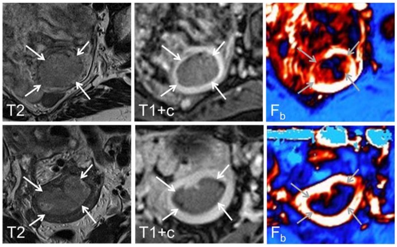
Figure 4.
Axial oblique T2-weighted MRI (left panel), contrast-enhanced T1-weighted MRI (middle panel) and parametric blood flow (Fb, right panel) map from a 68 year-old patient with FIGO stage 1B, grade 3 and high tumour Fb (upper panel) and a 52 year-old patient with FIGO stage 1B, grade 1 and low tumour Fb (lower panel). At conventional diagnostic MRI the endometrial carcinomas (arrows) are typically depicted as hyper-intense relative to the surrounding myometrium on T2-weigthed image while hypo-intense on contrast-enhanced T1-weighed image. On the corresponding parametric maps high blood flow is depicted as white/red while low blood flow is depicted as dark blue.

