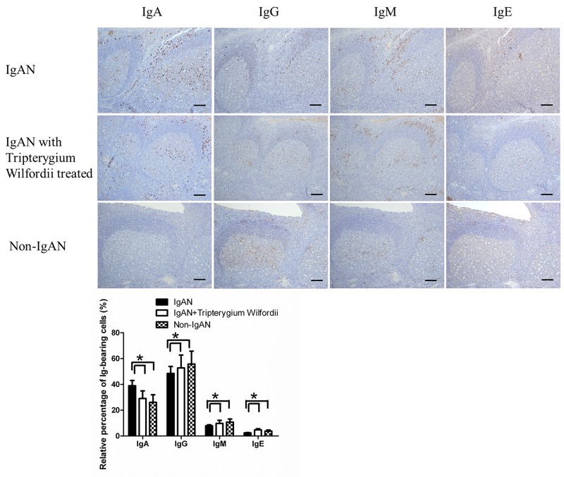
Figure 2. Among immunoglobulin classes, IgA was decreased in the tonsils of IgAN patients with Tripterygium Wilfordii treatment.
Immunohistochemistry on serial sections was used to show the expression of IgA, IgG, IgM and IgE in the tonsils. Bars, 500 μm. GC, germinal center. Positive cells were counted in low-power (100× magnification) fields for each patient. The slides were analyzed in blinded manner by two independent investigators. n = 20 for IgAN patients with Tripterygium Wilfordii treatment, n = 20 for IgAN patients without treatment and n = 20 for non-IgAN patients with chronic tonsillitis. Error bars indicate SEMs. *, P < 0.05 (Mann-Whitney U test).

