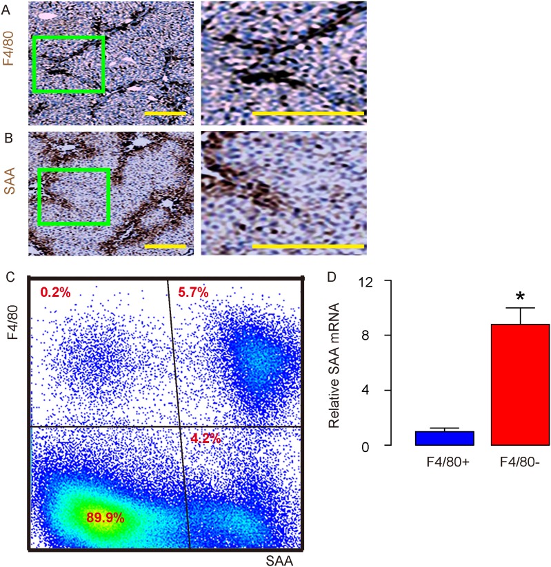
Figure 1. Macrophages are the major SAA-binding cells in the injured liver.
(A-B) Immunohistochemistry for F4/80 (A) and SAA (B) in mouse liver 8 weeks after carbon tetrachloride (CCl4) treatment. (C) Representative flow chart for F4/80 and SAA in dissociated mouse liver 8 weeks after CCl4 treatment. (D) RT-qPCR for SAA mRNA in purified F4/80+ vs F4/80- cells from dissociated mouse liver 8 weeks after CCl4 treatment. *p<0.05. n=5. Scale bars are 50 μm.

