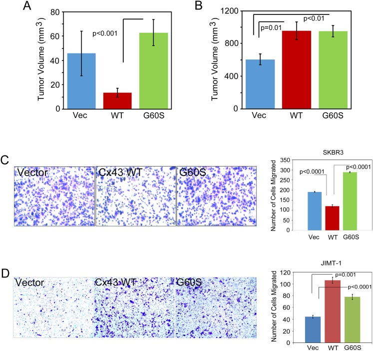
Figure 4. Expression of Cx43 in JIMT-1 cells promotes tumor growth and cell migration.
(A) SK-BR-3 expressing vector control, Cx43, or Cx43 G60S were injected into the mammary fat pad of immunocompromised mice to assess mammary tumor xenograft formation and growth. Tumor volume analysis on day 32 post-injection is presented. Student’s T-test was performed to determine p-values, p<0.001 as indicated, n=10 animals per group. (B) JIMT-1 expressing vector control or Cx43 were injected into the mammary fat pad of immunocompromised mice to assess mammary tumor xenograft formation and growth. Tumor volume analysis on day 32 post-injection is presented. Student’s T-test was performed to determine p-values, p= or < 0.01 as indicated, n=10 animals per group. (C) SK-BR-3 cells expressing vector control, Cx43, or Cx43 G60S were assessed by transwell assay to evaluate migration. Representative images show that Cx43 expressing cells migrate to a lesser extent than control or Cx43 G60S cells. Number of cells migrated was quantitated and students T-test was used to determine p-values, p<0.0001 as indicated, n= 4 samples per experiment. (D) JIMT-1 cells expressing vector control, Cx43, or Cx43 G60S were assessed by transwell assay to evaluate migration. Representative images show that Cx43 and Cx43 G60S expressing cells migrate to a greater extent than control cells. Number of cells migrated was quantitated and students T-test was used to determine p-values, p<0.0001 as indicated, n= 4 samples per experiment.

