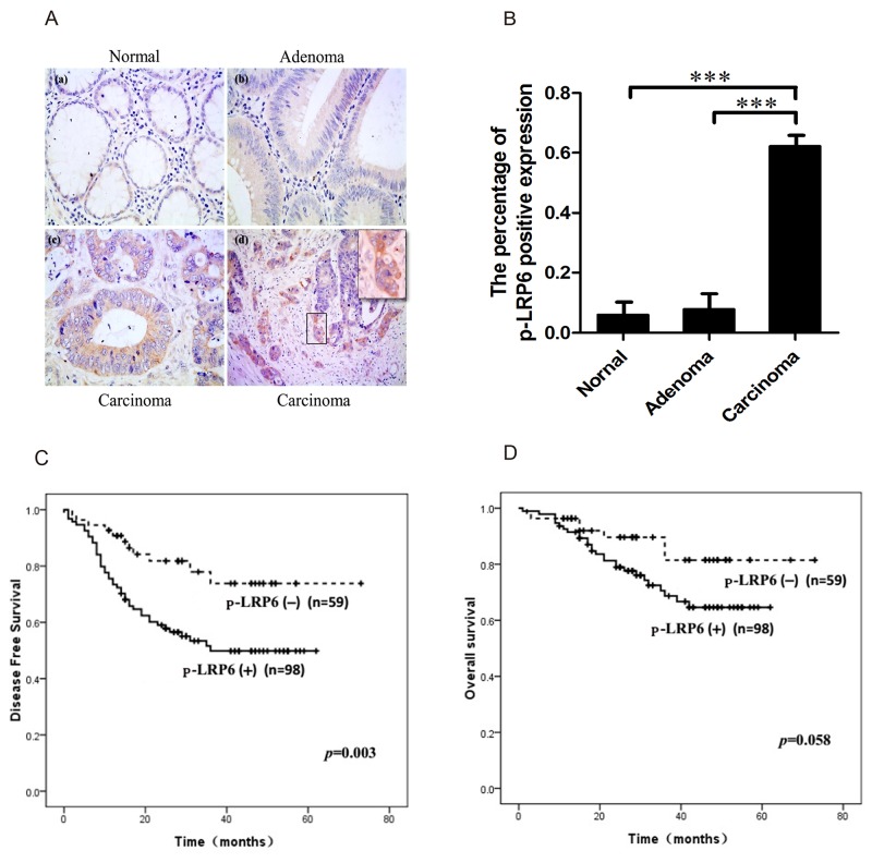
Figure 1. Immuno-staining of p-LRP6 correlated with outcome of colorectal cancers.
(A) The representative images of p-LRP6 staining in normal glandular cells (a), adenoma (b) and carcinoma (c) (×200). High magnification of p-LRP6 stained in front zone of cancer invasion (×800) (d). (B) The percentage of positive p-LRP6 expression in normal glandular cells, adenoma, and colorectal carcinoma. (C) Kaplan-Meier survival curves for the relation of p-LRP6 immuno-staining with DFS. (D) Kaplan-Meier survival curves for the relation of p-LRP6 immuno-staining with OS. * p< 0.05, **p< 0.01, ***p< 0.001.

