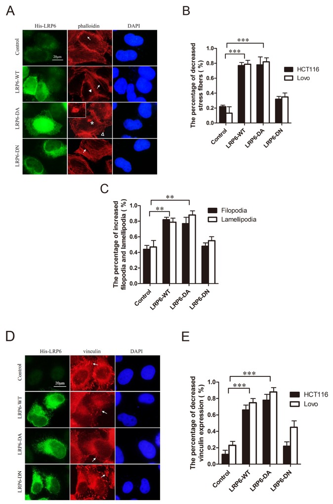
Figure 3. Activation of LRP6 increases actin remodeling.
(A) Overexpression of LRP6 induced actin remodeling. Lovo and HCT116 cells were transfected by control vector, LRP6-WT, LRP6-DA and LRP6-DN and doubly stained by His and phalloidin. Arrows indicate stress fibers, asterisk indicate the formation of invadopodia, arrowhead indicate filopodia and hollow arrowhead indicate lamellipodia. The data showed was from HCT116 cells. (B) The percentage of decreased stress fibers in each group with statistical analysis. (C) The percentage of increased filopodia and lamellipodia in each group with statistical analysis on HCT116 cells. (D) Overexpression of LRP6 reduced focal adhesions of cells. Lovo and HCT116 cells were transfected by control vector, LRP6-WT, LRP6-DA and LRP6-DN and doubly stained by His and vinculin. The arrows indicate focal adhesions. The data showed was from HCT116 cells. (E) The percentage of decreased vinculin expression in each group with statistical analysis. Scale bar, 20 μm. Values represent mean ± SD of three experiments. * p< 0.05, **p< 0.01, ***p< 0.001. All the experiments were repeated at least three times.

