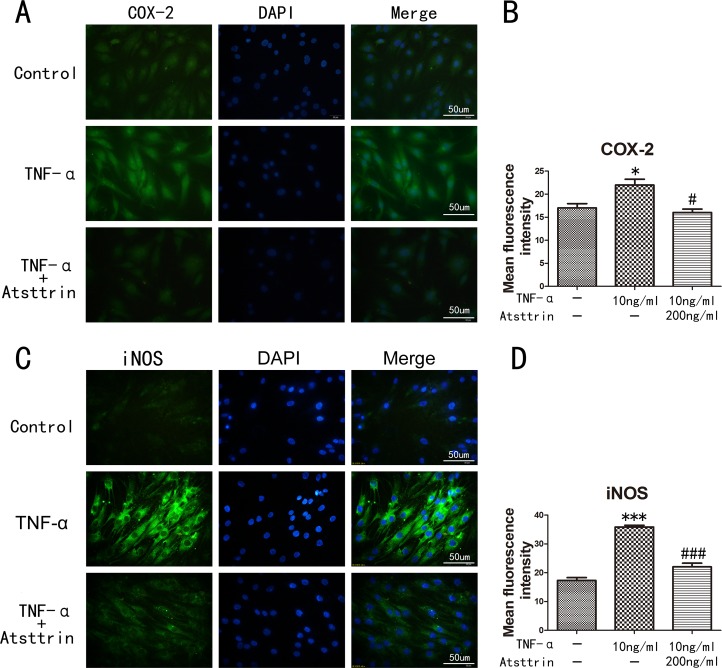
Figure 4. Inflammatory cytokines in human nucleus cells detected by immunofluorescent staining.
(A, C) Expression of iNOS and COX-2 in human NP cells detected by the immunofluorescence. (B, D) Analysis of the mean fluorescence intensity of COX-2 and iNOS according to the result of immunofluorescence results. The NP cells was treated with TNF-α (10ng/ml) in the presence or absence of Atsttrin (200 ng/ml), scal bars=50 um. *P<0.05, ***P<0.001, VS control group. #P<0.05, ###P<0.001, VS TNF-α treatment group.

