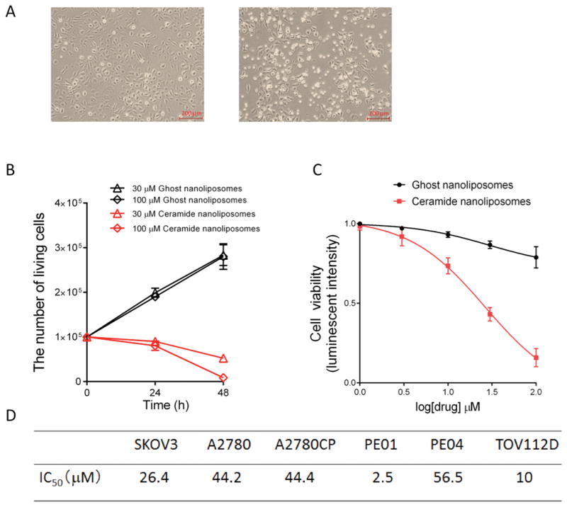
Figure 1. Cytotoxic effects of ceramide nanoliposomes in ovarian cancer cells.

A, SKOV3 cells (1 × 10⁵/well) were treated with 30 μM ceramide or ghost nanoliposomes for 24 h and then imaged by a phase-contrast microscopy. B, SKOV3 cells were treated with 30 or 100 μM nanoliposomes up to 48 h. The number of living cells was counted. The data represent the mean ± SD (n = 3). C, SKOV3 cells were treated with 1, 3, 10, 30, or 100 μM nanoliposomes. The cell viability was determined using a CellTiter-Glo luminescent cell viability assay according to the manufacturer’s protocol. The results are expressed as the percentages of 1 μM ghost nanoliposomes and the data represent the mean ± SD (n = 3). D, IC50 values were determined by GraphPad prism. The IC50 values of ceramide nanoliposomes in six kinds of ovarian cancer cell lines were shown.
