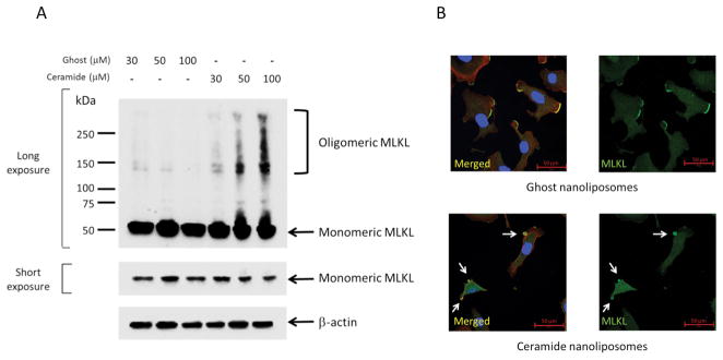
Figure 3. Activation of MLKL by ceramide nanoliposomes.
A, SKOV3 cells were transfected with MLKL-V5 vectors for 24 h followed by treatment with the indicated concentrations of ceramide or ghost nanoliposomes for 24 h. Cellular proteins extracted without reducing reagents were subjected to SDS-PAGE. Immunoblotting was performed using antibodies against V5 and β-actin. Three independent experiments were performed. Representative images are shown. B, SKOV3 cells were incubated with 30 μM ceramide or ghost nanoliposomes for 6 h and then were fixed followed by staining with TRITC-conjugated phalloidin (red), Hoechst 33342 (blue), MLKL (green). Imaging was performed by confocal microscopy, and representative images are shown. Arrows show the blebbing membranes.

