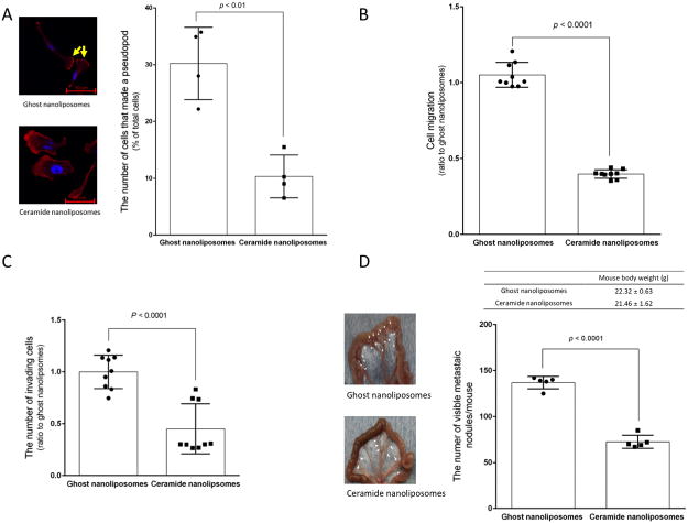
Figure 6. Effects of ceramide nanoliposomes on metastatic growth.
A, SKOV3 cells were treated with 30 μM ceramide or ghost nanoliposomes for 6 h. Cells were fixed and counterstained with TRITC-conjugated phalloidin (red) and Hoechst 33342 (blue). Imaging was performed by confocal microscopy and the cell number forming lamellipodia were determined by counting more than 300 cells. The values represent the percentage of cells forming lamellipodia relative to total cells and the data represent the mean ± SD (n = 4). Four independent experiments were performed and yellow arrows show lamellipodia. Statistical analyses were performed by unpaired, student t-test. B and C, SKOV3 cells were treated with 30 μM ceramide nanoliposomes or ghost nanoliposomes for 18 h and then the assay for migration (B) and invasion (C) was performed as described in “Materials and Methods”. Data are represented as the percentage compared with ghost nanoliposomes’ group. The data represent are the means ± SD (n = 9). Statistical analyses were performed by unpaired, Student t-test. D, SKOV3 cells (5 × 10⁶ / mouse) were injected intraperitoneally into peritoneal cavity of 4 weeks-old female nude mice. One day after implantation of cells, mice were intraperitoneally treated with ceramide or ghost nanoliposomes (40 mg/kg/day) continuously for 3 days. Four weeks later after inoculation, mice were euthanized to determine metastatic growth in the mesentery. The number of metastatic nodules was determined and mouse body weight was measured (n = 5). The data represent the mean ± SD and the representative images of specimens from mice were shown. White arrowheads indicate metastatic nodules. Statistical analyses were performed by unpaired, student t-test.

