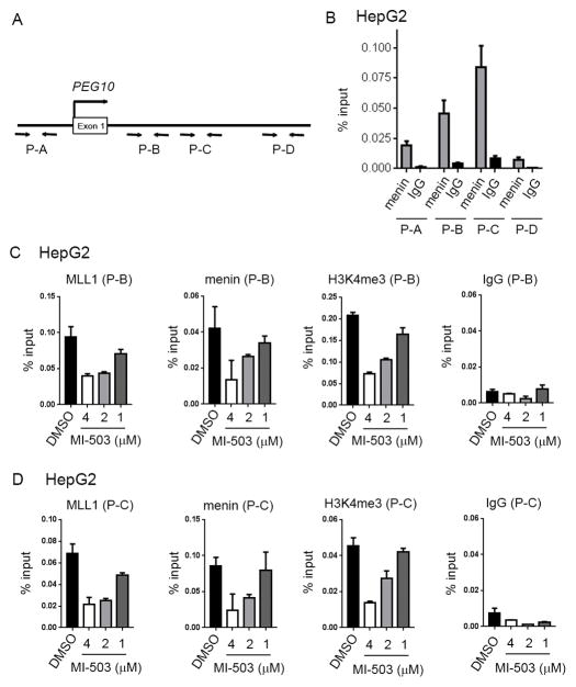
Figure 4. Epigenetic regulation of PEG10 by menin-MLL1 in HCC.
A. Diagram showing the location of primers at the promoter and downstream regions of PEG10. Genomic coordinates are provided in Supplementary Methods. B. Menin binding to different regions (P-A – P-D) of PEG10 obtained from the chromatin immunoprecipitation (ChiP) experiment performed in HepG2 cells. C, D. ChiP experiments performed in HepG2 cells upon 6 days of treatment with various concentrations of MI-503 or DMSO to detect menin and MLL1 binding as well as H3K4me3 on PEG10. Real-time PCR was performed on the precipitated DNAs with P-B (C) and P-C (D) primers for PEG10 genomic regions. IgG was used as a control. Data represent mean of triplicates ± SD.

