
Figure 1. PLX8394 effectively reduces ERK1/2 signaling and tumor volume in vivo.
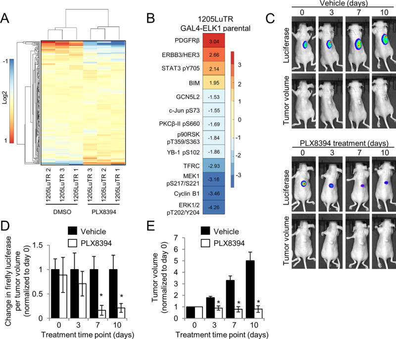
A. 1205LuTR GAL4-ELK1 cells were treated for 24 hours with either DMSO or 0.5 μM PLX8394. Lysates were obtained from three independent experiments and processed for RPPA analysis. A heat map was generated using median-centered data across each protein measurement for each sample. B. Proteins with a p value ≤ 0.01 and a fold change of ≥ 1.5 that were significantly altered following PLX8394 treatment. C. Mice bearing 1205LuTR GAL4-ELK1 xenografts were fed either vehicle chow or PLX8394 laced chow. Representative images of a vehicle and PLX8394 treated mouse with overlaid luciferase output across 10 days of treatment are shown. D. Quantification of firefly luciferase. Graph depicts fold change in luciferase output per tumor volume compared to vehicle for each day of treatment. E. Average fold change in tumor volume between mice fed vehicle chow and PLX8394-laced chow.