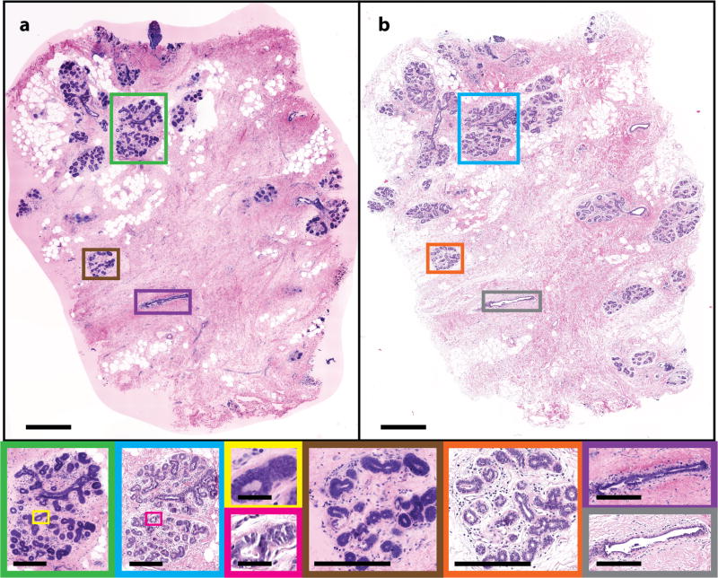
Fig. 2.
Normal human breast tissue. (a) NLM and (b) FFPE H&E histology images (500 µm scale bar). Normal TDLUs (NLM: green and brown box; FFPE H&E: blue and orange box) (250 µm scale bar), individual acini (NLM: yellow box; FFPE H&E: pink box) (50 µm scale bar) and larger ducts (NLM: brown box; FFPE H&E: gray box) (250 µm scale bar) are shown magnified. NLM: https://slide-atlas.org/link/zmhbwg. FFPE H&E: https://slide-atlas.org/link/noazj9.

