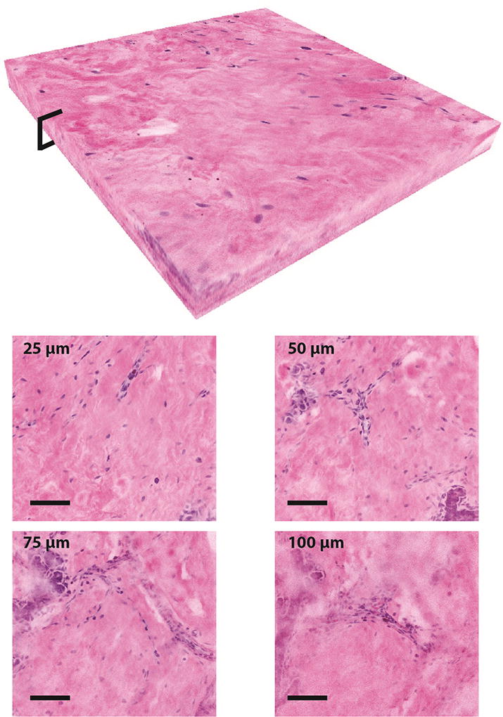
Fig. 5.

A 500×500×100 µm three-dimensional stack of NLM images demonstrating imaging at different depths in normal breast tissue. The volumetric reconstruction shows image data from the surface to 100 µm deep with digital gain applied to the images in post processing to match the average frame intensity without changing incident laser power or detector gain (scalebars: 100 µm). Selected NLM images from 25 µm, 50 µm, 75 µm, and 100 µm depths are shown.
