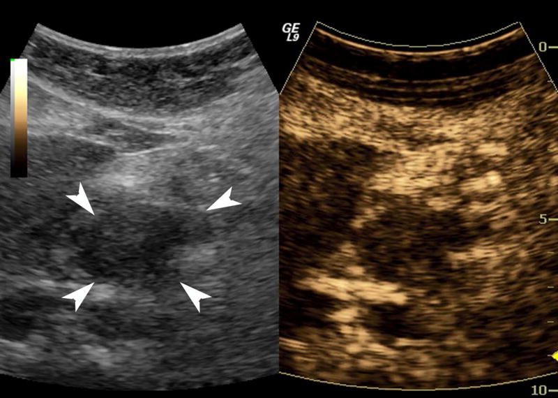
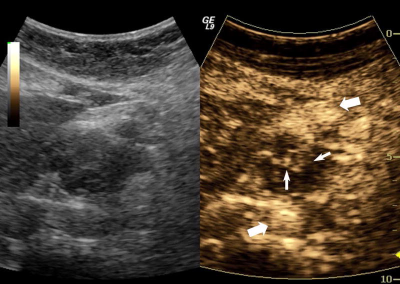
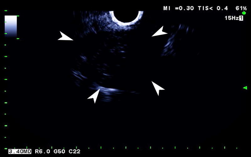
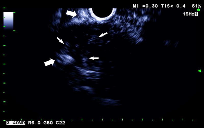
Figure 1.
A pancreatic adenocarcinoma (arrowheads) scanned in dual grayscale/SHI mode (SHI is depicted in golden hues on the right) before (a) and after (b) injection of Definity. The size of this tumor was approximately 3.0×4.0×4.4 cm. The same lesion depicted in contrast EUS mode before (c) and after (d) contrast administration. Notice, the larger vessels (large arrows) external to the cancer seen with both EUS and SHI, while smaller neovessels inside the mass are more prominent in SHI mode (small arrows).


