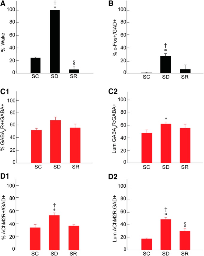
Figure 2.
Sleep-wake states, c-Fos, GABAA, and AChM2 receptors in RFMes GABAergic neurons across groups. A, The percentage of time spent in wake during the 2 h preceding termination differed significantly across groups, being higher in SD as compared to SC and SR and lower in SR as compared to SC. B, The % of GAD+ neurons that were positively immunostained for c-Fos (+) differed significantly between groups, being greater in SD as compared to SC and SR. C, The % of GABA+ neurons which were positively immunostained for the GABAAR (+) increased insignificantly following SD as compared to SC and SR (C1). The luminance of the GABAAR immunofluorescence on GABAAR+/GABA+ neurons differed significantly, being higher in SD as compared to SC (C2). D, The % of GAD+ neurons which were positively immunostained for AChM2R (+) differed significantly between groups, being higher in SD as compared to SC and SR (D1). The luminance of the AChM2R immunofluorescence on AChM2R+/GAD+ neurons differed significantly, being higher in SD as compared to SC and SR (D2). Note that the changes in GABARs and AChM2Rs on RFMes GABAergic neurons parallel the changes in % Wake and % c-Fos+/GAD+ across groups; * indicates significant difference of SD relative to SC; † indicates significant difference of SD relative to SR; § indicates significant difference of SR relative to SC (p < 0.05), according to post hoc paired comparisons following one-way ANOVA (Table 1).

