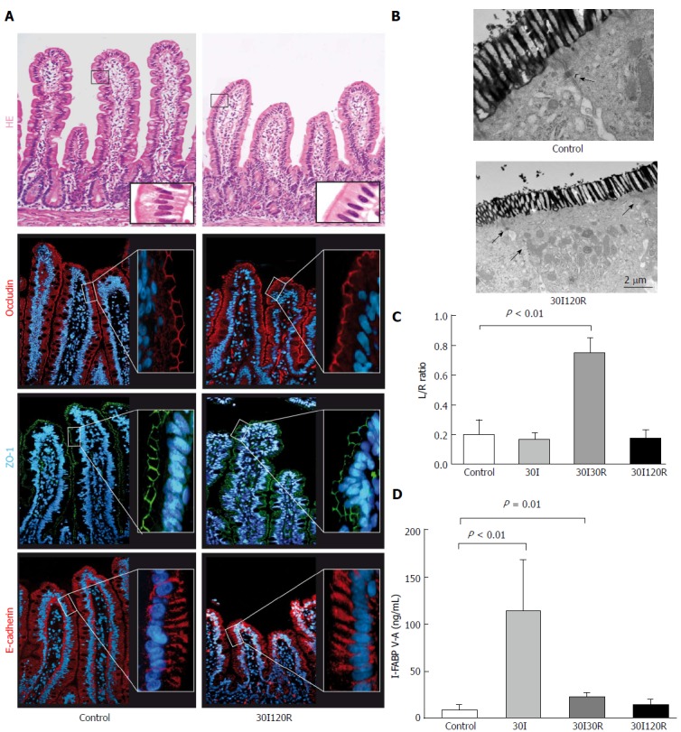
Figure 4.

The intestinal barrier is restored after 30 min of ischema followed by 120 min of reperfusion. A: At 120 min of reperfusion (30I120R) the epithelial lining appeared histologically intact compared to control tissue with ZO-1, occludin and E-Cadherin normally distributed across the villi.); B: In addition, electron microscopy revealed that lanthanum was no longer present in the paracellular spaces, indicating restored tight junction integrity; C: Moreover, the observed restoration of the intestinal barrier seems to correlate with permeability with the plasma L/R ratio normalized to 0.17 ± 0.06 which was no longer significantly different from control; D: Plasma I-FABP levels, reflecting intestinal epithelial damage, also returned to 14.77 ng/mL ± 5.46 ng/mL and were no longer significantly elevated compared to control. Electron microscopy scale bars = 0.5 μm and 2 μm respectively.
