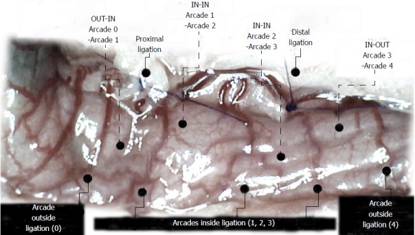
Figure 1.

Assessment of arcade vessels arcade 0; arcade 1, 2, 3; arcade 4 (dorsal colon side before the initiation of therapy, USB microscope camera). The ligations include three major arcades within the ligations (arcade 1, 2, 3), and thereby the possibility of bypassing both obstructions caused by the proximal and distal ligations (full lines) by an additional network of vessels located between the arcades (outside (arcade 0, arcade 4) and inside ligation (arcade 1, 2, 3); outside (arcade 0)-inside arcade 1); inside -inside(arcade 1-arcade 2; arcade 2-arcade 3); inside (arcade 3)-outside (arcade 4) (dashed lines) at the both ventral and dorsal sides of the colon. Note that there is no contact between inside arcade 3 and outside arcade 4. The appearance of the arcade vessels at specific time points [A (1 minute before therapy), B (next 5 min), C (the next 5 minutes, until the 10th min), D (until the end of the 15th min)] was assessed between (1) the last arcade proximal to first ligation (arcade 0) at the first arcade distal to first ligation, inside the ligated segment (arcade 1) (OUT-IN) (0-1, Figure 2, ♦--, Figures 5, 6, 13, 14); (2) between the next arcade (arcade 2), middle arcade inside ligations (IN-IN) (1-2, Figure 2; ▲, Figures 5, 6, 13, 14) and the following arcade (2-3, Figure 2; --X--, Figures 5, 6, 13, 14), and finally presentation to the first outside arcade distal to the second ligation (arcade 4) (IN-OUT) (3-4, Figure 2; ■, Figures 5, 6, 13, 14).
