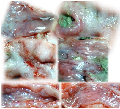
Figure 20.

IC + OB rats underwent additional colon obstruction for three days; the colon was then opened before sacrifice on day 10 [i.e., one week after the additional colon obstruction had been removed and bath therapy applied (saline, upper and middle; BPC 157, lower)]. The examination revealed progressive worsening in the saline-treated animals (upper and middle) and recovery in the BPC 157-treated animals (lower) (see Figure 11). The control rats exhibited extremely large pale areas without mucosal folds covering the entire area between ligations (middle); such areas were present even beyond the area of the ligation (upper), together with ulcerations in the deprived, apparently enlarged colon segment (middle). BPC 157-treated rats exhibited almost completely spared mucosa (very small pale areas) and no ulceration; the previously ligated colon segment was of normal diameter (lower). The images were obtained using a USB microscope camera.
