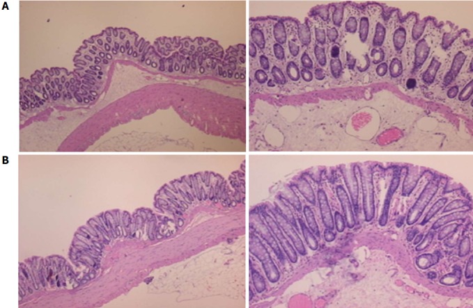
Figure 23.

Characteristic microscopic appearance of the colon in IC (A, control) rats and BP 157-treated IC rats (B) 15 min after ligation. HE, 4 × objective (left) and 10 × objective (right). A: Severe edema of the lamina propria and continuous diffuse edema of the submucosa can be observed. Pronounced dilatation and stasis of the submucosal blood vessels is also present. B: Mild edema of the lamina propria and focally present mild-to-intermediate edema of the submucosa can be observed. Although stasis of the submucosal blood vessels is present, the dilatation of the veins appears to be less pronounced.
