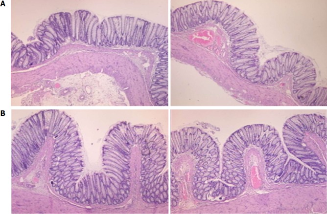
Figure 25.

Microscopic appearance of the colon in IC + OB rats (A, control) and BPC 157-treated IC + OB rats (B) after additional colon obstruction for three days one week after the additional colon obstruction was removed. Control (saline bath); 10 d; BPC 157, 10 d. HE, 4 × objective (left); 10 × objective (right). A: Mild edema of the lamina propria and diffuse mild-to-intermediate edema of the submucosa, along with the formation of collagen, can be observed. Stasis of the submucosal blood vessels is also present. The rugae are broadened and flattened, so the mucosa appears flattened macroscopically. B: Mild edema of the lamina propria and practically no edema of the submucosa is observed. Stasis of the submucosal blood vessels is also present but to a much lesser extent than in the controls. The rugae are histologically well formed and only occasionally slightly broadened. There is minimal formation of new collagen fibers in the submucosa.
