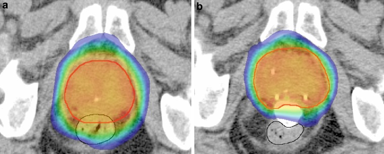
Fig. 1.
Color-wash isodose distribution projected on an axial CT slice before (a) and after RBI implantation (b) in the same patient with the planning target volume in red. Without RBI (a), the high-dose region >80% (green isodose) overlaps with the entire ventral part of the rectum (black line), whereas with the RBI in situ (b) the rectum is exposed to a dose <65% (blue isodose)

