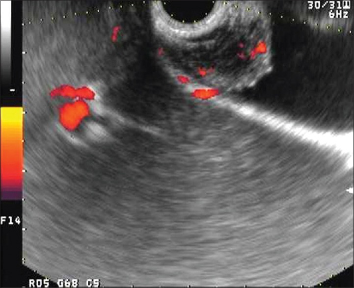
Figure 1.

Endoscopic ultrasonography image showing a well-defined hypoechoic heterogeneous mass with a hyperechoic-layered structure arising from the gallbladder wall. Power Doppler endoscopic ultrasonography revealed spotty increased flow on the tumor
