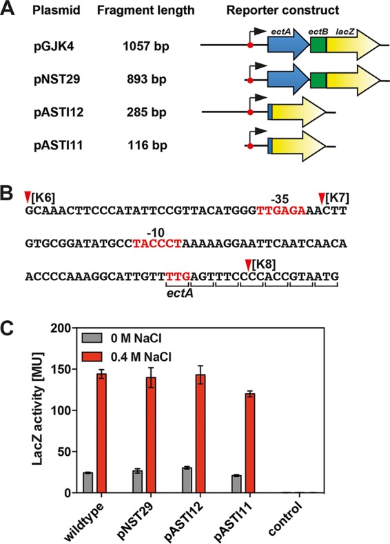
FIG 4.

A minimal DNA fragment directing osmoregulated ect transcription. (A) Physical structures of ect-lacZ reporter constructs. (B) DNA sequence of the DNA fragment present in plasmid pASTI11. The deletion endpoints K6, K7, and K8 defined in the experiments for Fig. 3A are indicated, and the −35 and −10 elements of the ect promoter are highlighted. The fusion junction within ectA to the lacZ reporter gene lies in codon seven. (C) β-Galactosidase reporter enzyme activity in cells of strain MC4100 carrying the plasmids depicted in panel A. Cells of MC4100 carrying the vector (pBBR1MCS-2-lacZ) used to construct plasmids pGJK4, pNST29, pASTI12, and pASTI11 were used as the control. Cultures were grown in MMA or MMA containing 0.4 M NaCl and were harvested and processed for β-galactosidase enzyme activity when they reached an optical density (OD578) of about 1.8. The data shown were derived from four independently grown cultures, and each enzyme assay was performed at least twice. β-Galactosidase enzyme activity is given in Miller units (MU) (108).
