
Figure 2.
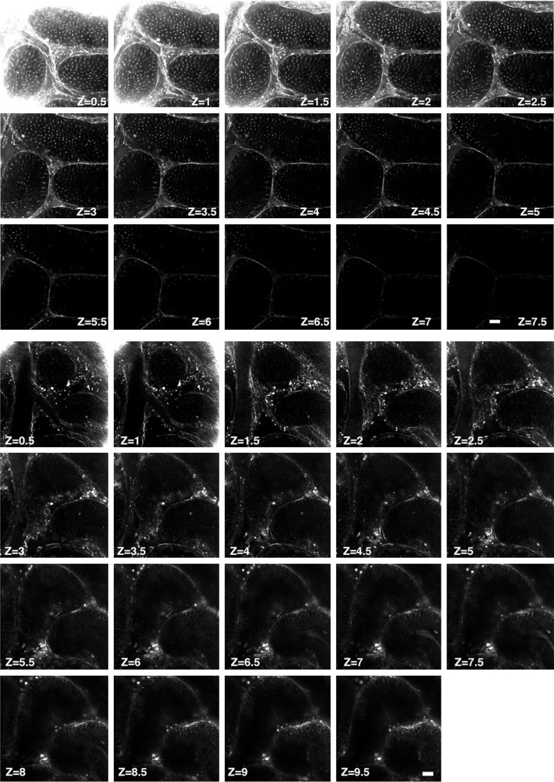
Second-harmonic generation microcopy imaging of germ cells in testicular tubules in normal mouse testes. Upper panel: Illustrating the second-harmonic microscopy imaging (SHIM) finding for identification of living normal mouse testes. This second-harmonic generation (SHG) optical sections clearly and consecutively showed the microtubule emitted signals in the seminiferous tubules from superficial to deeper areas. Zone (Z) = 0.5, indicated 0.5 μM and so on, Z = 7.5, indicated 7.5 μM. Lower panel: Illustrating second-harmonic microscopy imaging (SHIM) finding for identification of living doxorubicin-treated mouse testes. This second-harmonic generation (SHG) optical sections clearly and consecutively showed the microtubule emitted signals in the seminiferous tubules from superficial to more deeper areas. As expected, 15 mg/kg cumulative dose doxorubicin, the seminiferous tubules exhibited much fewer SHG signals in each cross-section area. Zone (Z) = 0.5, indicated 0.5 μM and so on, Z = 7.5, indicated 7.5 μM.