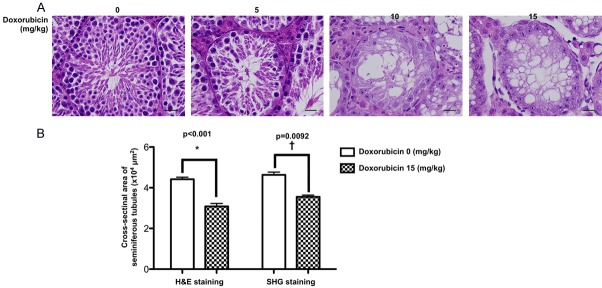
Figure 3.
Histopathological finding of mouse testicular structure without and with doxorubicin treatment. A. In normal control mouse (i.e., 0 mg/kg Doxo), the microscopy clearly identified the germ cells organized in concentric layers constituting the seminiferous epithelium, tubular lumen, interstitial tissue, interstitial cells in the tubular spaces, spermatogenic lineages and the sertoli cells in seminiferous tubules of testis. On the other hand, the testicular tubules in mouse treated with doxorubicin (5 mg/kg) were identified to have smaller seminiferous tubules and fewer germs cells in the seminiferous tubules than normal control. Additionally, in mouse treated with a higher dose of doxorubicin (10 mg/kg and 15 mg/kg), the testicular tubules were found to be almost disappeared with disorganized seminiferous epithelium. Furthermore, the seminiferous tubules contained only sertoli cells without germ cells. Besides, vacuolization was detected in the seminiferous tubules from 10 mg/kg and 15 mg/kg doxorubicin-treated mice. B. Left bar chart: analytical result of cross-sectional areas of seminiferous tubules assessed by H.E., stain, normal vs. Doxo-treated, *indicated p = 0.0092. Right bar chart: analytical results of cross-sectional areas of seminiferous tubules assessed by SHIM imaging, normal vs. Doxo-treated, †indicated p<0.001. Data represent the mean of 10 random circular cross-sections ± standard error of means (SEM). n = 6 in each group. Doxo = doxorubicin

