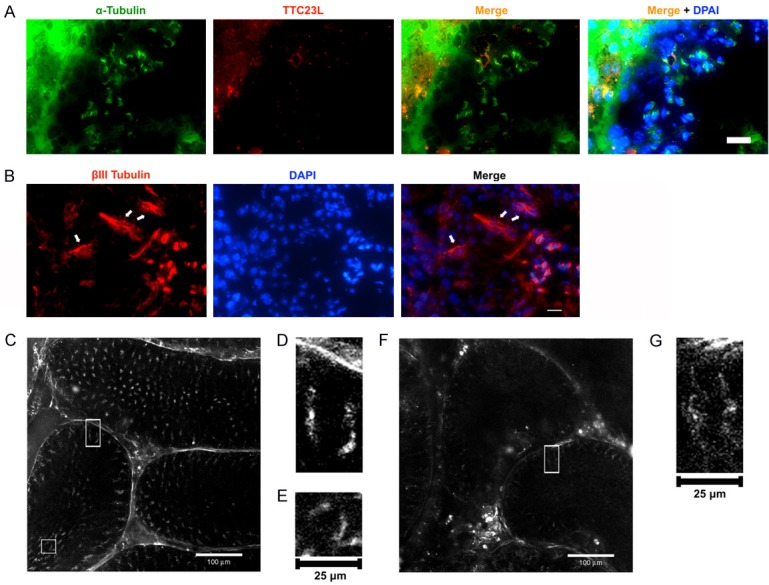
Figure 4.

Microtubule machettes in spermatids and axoplasmic microtubules in Sertoli cells, and SHIM imaging resolved microtubule structures in sertoli cell and spermatids cells. (A) Immunofluorescent microscopic finding showed the α-tubulin positive cells in testes. Testis cryosections were stained by α-tubulin (green) antibody and TTC23L (red) antibody, respectively. The α-tubulin staining indicated the microtubule machettes in spermatids and TTC23L staining showed a punctuated pattern in spermatids associated with microtubule manchattes. Blue color indicated nuclei stained by DAPI. (B) The anti-beta tubulin III antibody (green) was used as a marker for sertoli cells. The immunostaining of βIII-tubulin protein was restricted to Sertoli cells of the seminiferous epithelium. Arrows indicated βIII-tubulin antibody (red) staining. Blue color corresponded to nuclei stained by DAPI. Scale bars in right lower corner represent 60 µm. (C) SHIM imaging analysis at Z = 3.5 μm in testis without doxorubicin treatment displayed microtubule manchette in spermatids and axoplasmic microtubule in sertoli cells. Bar = 100 μm. (D) Rectangle in (C) Indicating the area to be selected for high-power magnification shows axoplasmic microtubules. (E) Square in (C) Indicating area to be selected for magnification. The high-power magnification of sub-area showed microtubule manchette. (F) A SHIM imaging analysis at Z = 1 μm in testis with doxorubicin treatment displayed only axoplsmic microtubules in sertoli cells to be observed and the microtubule machettes were absent. (G) Rectangle in (F) indicating area to be selected for magnification. The high-power magnification shows axoplasmic microtubules.
