Figure 5.

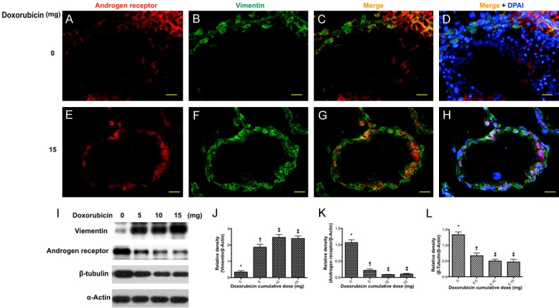
Immunostaining and immunoblot of vimentin in sertoli cells. Upper panel: showed the immunofluorescent (IF) findings in situation without doxorubicin treatment. (A and B) Illustrating the IF microscopic finding for identifying the immunostaining of androgen receptor (red) (A) and vimentin in seminiferous tubules (B), respectively. (C and D) Merge IF microscopic finding indicated the Vimentin-positive sertoli cells were present in the seminiferous epithelium. (D) Blue color indicated nuclei stained by DAPI. DAPI-positively stained spermatids and spermatozoa were richly distributed in the seminiferous tubules. Lower panel: showed the IF findings in situation with doxorubicin treatment. (E and F) Illustrating the immunostaining of androgen receptor (red) (E) and vimentin in seminiferous tubules (F), respectively. (G) The IF microscopic finding showed that vimentin-positive sertoli cells were present in the seminiferous epithelium. (H) Blue color indicated nuclei stained by DAPI. No DAPI-stained spermatids and spermatozoa were identified in the seminiferous tubules. Scale bars in right lower corner represent 20 µm. (I) Showing Western blot results of sertoli marker vimentin, androgen receptor and β-tubulin, respectively. (J) Protein expression of vimentin, * vs. other groups with different (†, ‡, p<0.001. (K) Protein expression of androgen receptor (AR), * vs. other groups with different (†, ‡, p<0.001. (L) Protein expression of β-tubulin, * vs. other groups with different (†, ‡, p<0.001. All statistical analyses were performed by one-way ANOVA, followed by Bonferroni multiple comparison post hoc test (n = 6 for each group). Symbols (*, †, ‡) indicate significance at the 0.05 level. Doxo = doxorubicin.
