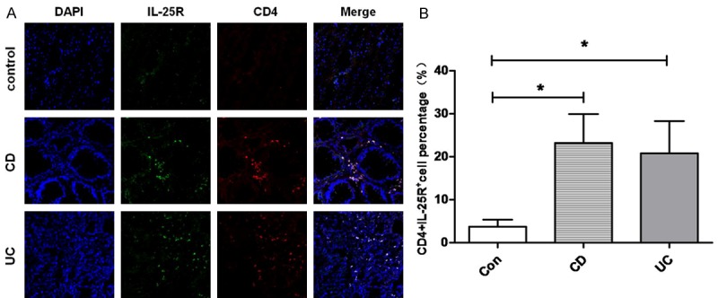
Figure 1.

Colocalization between IL-25R and CD4 in the intestinal mucosa. A: Representative images from inflamed mucosa of a CD patient, a UC patient, and a healthy control (×200). IL-25R+CD4+ cells were detected by double immunofluorescence staining. B: Quantification of IL-25R+CD4+ cells in the intestinal mucosa of healthy control, CD patient, and UC patient (*P<0.05 vs control group). Data are expressed as mean number of positive cells per high power field ± SEM from 3 independent experiments.
