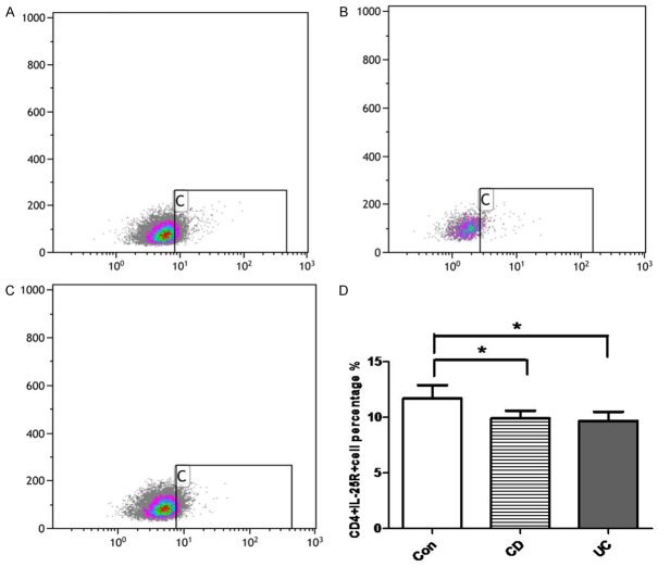
Figure 4.
Proportion of CD4+IL-25R+ cells in the peripheral blood. CD4+IL-25R+ cells in the peripheral blood of healthy controls (A), CD patients (B) and UC patients (C) were detected by flow cytometry. (D) Quantification of CD4+IL-25R+ cells in the intestinal mucosa of healthy control, CD patient, and UC patient. *P<0.05 vs healthy controls.

