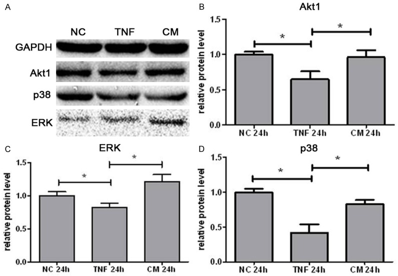
Figure 7.

Protein expression of AKT1 (B), ERK(C) and P38 (D) in IEC-6 cells after 24-h exposure to different media. Protein expression was detected by Western blotting. (A) Representative WB images were shown for NC groups, TNF group and CM group. Data are represented as means ± SEM from three independent experiments (*P<0.05). NC: negative control group; TNF: control medium group; CM: IL-25 primed MSCs medium group.
