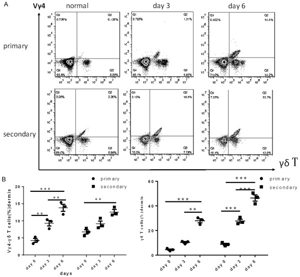
Figure 2.
The number of Vγ4 +γδ T cells in dermal tissue. A. Vγ4 +γδ T cells were measured in a suspension of digested dermal cells from mice with psoriasis-like skin inflammation on day 0, 3 and 6, gated on total T cells. B. The percent of Vγ4 -γδ T cells and γδ T cells on different days. The values were calculated as the mean ± SEM (n=3), **p<0.01, ***p<0.001.

