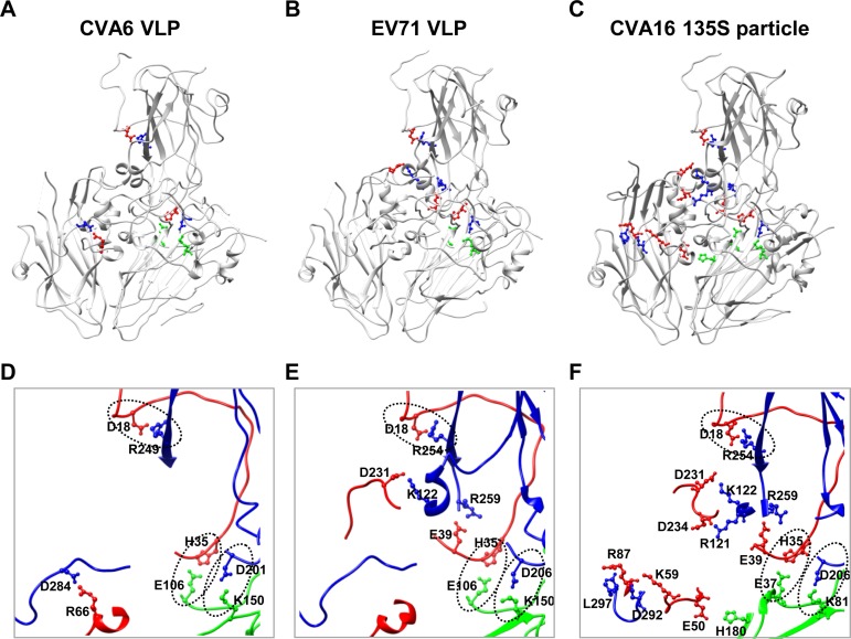
FIG 5.
Salt bridges within the protomers of the CVA6 VLP, EV71 VLP, and CVA16 135S particle. (A to C) Salt bridge locations within the protomers of the CVA6 VLP (A), EV71 VLP (B), and CVA16 135S particle (C). Salt bridge-forming residues within VP1, VP0/VP2 (VP0 for the CVA6 VLP and EV71 VLP and VP2 for the CVA16 135S particle), and VP3 are colored in blue, green, and red, respectively, while the other portions of the proteins are in gray. (D to F) Zoom-in view of the salt bridge regions enlarged from panels A, B, and C, respectively. The three common salt bridges observed in the CVA6 VLP, EV71 VLP, and CVA16 135S particle are indicated by oval dashed lines.

