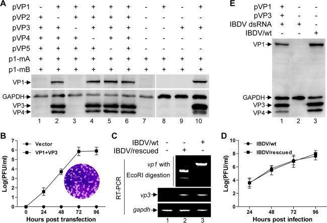
FIG 2.
VP1 and VP3 are the minimal viral components required and sufficient for rescue of infectious virus from the uncapped IBDV RNA. (A) HEK 293T cells were cotransfected with p1-mA and p1-mB together with different combinations of pVP1, pVP2, pVP3, pVP4, and pVP5 as indicated. The supernatant of transfected cells was collected at 72 h posttransfection and passaged on DF-1 cells, and subsequently lysates of passaged cells were subjected to Western blot analysis using antibodies against VP1, VP3, and VP4. Anti-GAPDH was used as a loading control. (B) HEK 293T cells were cotransfected with p1-mA and p1-mB together with pVP1 and pVP3 or pCI-neo empty plasmid. Supernatant of the transfected cells was collected at the indicated time points posttransfection, and the viral titer was measured by plaque assay. The curve was plotted based on three independent experiments, and values are expressed as means ± standard deviations. The inset shows an example of plaques formed on a monolayer of DF-1 cells inoculated with the supernatant from HEK 293T cells cotransfected with p1-mA, p1-mB, pVP1, and pVP3 for 72 h. (C) RT-PCR analysis of vp1 and vp3 for validation of the viral RNA transcription level in the infected DF-1 cells, and discrimination of the rescued virus (IBDV/rescued) from wild-type virus (IBDV/wt) by EcoRI digestion analysis of the amplified vp1 gene fragment. (D) The plaque assay was performed to establish the one-step growth curves of the rescued viruses (IBDV/rescued) and the wild-type viruses (IBDV/wt) with the inoculation of virus at a multiplicity of infection of 0.1. (E) HEK 293T cells were cotransfected with the purified IBDV genomic dsRNA together with pVP1 and pVP3 or pCI-neo empty plasmid. The supernatant was collected at 72 h posttransfection and passaged on DF-1 cells. Western blotting was performed to verify the rescue of infectious viruses using antibodies against VP1, VP3, and VP4.

