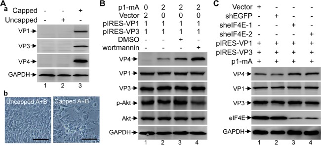
FIG 5.
VP1- and VP3-mediated translation initiation of uncapped plus-sense IBDV RNA transcripts is independent of the 5′ cap and the cap-binding protein eIF4E. (A) Western blot analysis of the cell lysate from HEK 293T cells transfected with the in vitro-transcribed IBDV RNAs of both A and B segments with or without an artificial addition of a 5′ cap, using antibodies against VP1, VP3, and VP4 (panel a). The supernatant was passaged at 72 h posttransfection, and the images were taken under a phase-contrast microscope at 12 h postinfection (panel b). Scale bar, 100 μm. (B) HEK 293T cells were transfected with the indicated plasmids, and the amount (in micrograms) of each plasmid for transfection is indicated above the lanes. The transfected cells were either untreated, mock (DMSO) treated (4 μl/well), or treated with wortmannin (800 nmol in 4 μl of DMSO/well) for 72 h. Western blot analysis was performed to analyze the expression of VP4, VP1, VP3, phospho-Akt (Ser473) (p-Akt), and total Akt. (C) HEK 293T cells were transfected with the shRNA plasmids for 24 h, and the resultant cells were cotransfected with p1-mA, pIRES-VP1, and pIRES-VP3 for another 72 h; Western blotting was performed to analyze the levels of VP4, VP1, VP3, and eIF4E.

