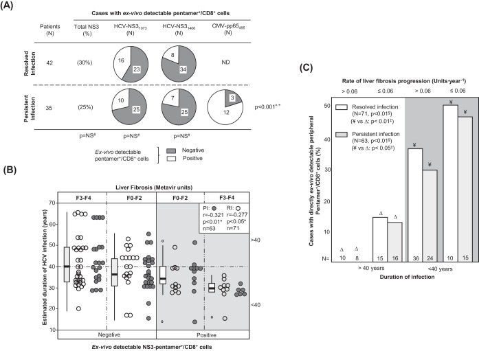
FIG 1.
Ex vivo detection of pentamer+/CD8+ cells. (A) Frequency of patients with resolved and persistent infections and HCV NS31073-, HCV NS31406-, and CMV pp65495-specific pentamer+/CD8+ cells detectable ex vivo. (B) Correlation between HCV infection length and detection of peripheral HCV pentamer+/CD8+ cells ex vivo, unbundled according to liver fibrosis stage. Box plots represent the distribution of the infection duration in each category when the data for patients with persistent infection and resolved infection after treatment are taken together. (C) Frequency of cases with peripheral pentamer+/CD8+ cells detectable ex vivo in relation to the duration of HCV infection (short/midduration, ≤40 years; long lasting, >40 years) and the rate of liver fibrosis progression. #, chi-square test; ¤, Spearman correlation test; ‡, Mann-Whitney U test; §, linear trend test; F, Metavir fibrosis stage; ND, not done; NS, nonsignificant; PI, persistent infection; RI, resolved infection after treatment; *, comparison between HCV-pentamer+/CD8+ and CMV-pentamer+/CD8+ cells in PI.

