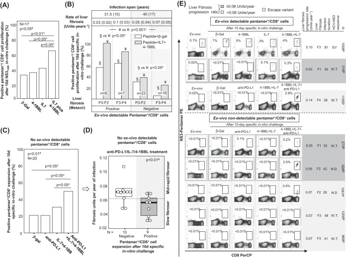
FIG 11.
Restoration of pentamer+/CD8+ cell reactivity. (A) In patients with persistent infection, the percentage of assays with pentamer+/CD8+ cell expansion after 10 days of NS31406 challenge in vitro in the presence of treatment with β-galactosidase (β-gal), IL-7, 4-1BBL, or IL-7 plus 4-1BBL. (B) Cases with pentamer+/CD8+ cell proliferation after antigen-specific stimulation in the presence of treatment with β-galactosidase or IL-7 plus 4-1BBL, according to the detection of peripheral pentamer+/CD8+ cells ex vivo. The rate of liver fibrosis progression and the duration of infection are expressed as the median plus interquartile range. Liver fibrosis is described by the Metavir score. (C) Percentage of cases without cells detectable ex vivo with pentamer+/CD8+ cell proliferation after treatment with β-galactosidase, anti-PD-L1, IL-7 plus 4-1BBL, or IL-7, 4-1BBL, and anti-PD-L1. (D) In patients with PI without cells detectable ex vivo, the liver fibrosis progression rate according to the proliferative potential after specific in vitro treatment with IL-7, 4-1BBL, and anti-PDL1. Box plots summarize the distribution of the liver fibrosis progression rate in each group. In the group positive for expansion, one case was not included because the estimated date of infection was unknown. (E) Representative dot plots showing the expansion of CD8+/pentamer+ cells in the presence of different treatment combinations, according to the detection of CD8+/pentamer+ cells ex vivo and the liver fibrosis progression rate. The data in each dot plot show the frequency of pentamer+/CD8+ cells out of the number of total CD8+ cells. EV, escape variant; F, Metavir fibrosis score; N.D., not done; ○, outlier; W.T.; wild type; #, Friedman test; γ, chi-square test; ¤, Wilcoxon test; &, Mann-Whitney U test.

