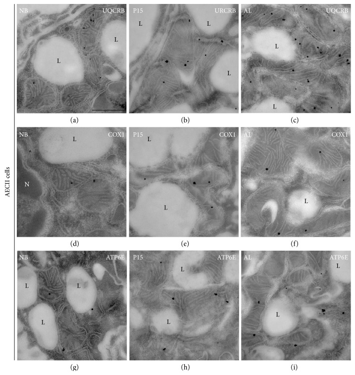
Figure 9.
Electron micrographs showing immunogold labelling for mitochondrial proteins in ultrathin sections of AECII in newborn (NB), P15, and adult (AL) animals. Lung tissue processed for immunoelectron microscopy was incubated with gold-labelled secondary antibody particles and thereafter contrasted with uranyl acetate and lead citrate prior to analysis by transmission electron microscopy. (a–i) Immunogold labelling in mitochondria of AECII for (a–c) complex III (UQCR2), (d, e) complex IV (COX1), and (g, h) complex V (ATP6E). S, secretory granule; L, lamellar bodies. Bars represent 0.5 μm.

