
Figure 6. Point mutations of PSD‐95 that disrupt calmodulin binding prevent BIC‐induced loss of PSD‐95 from spines.
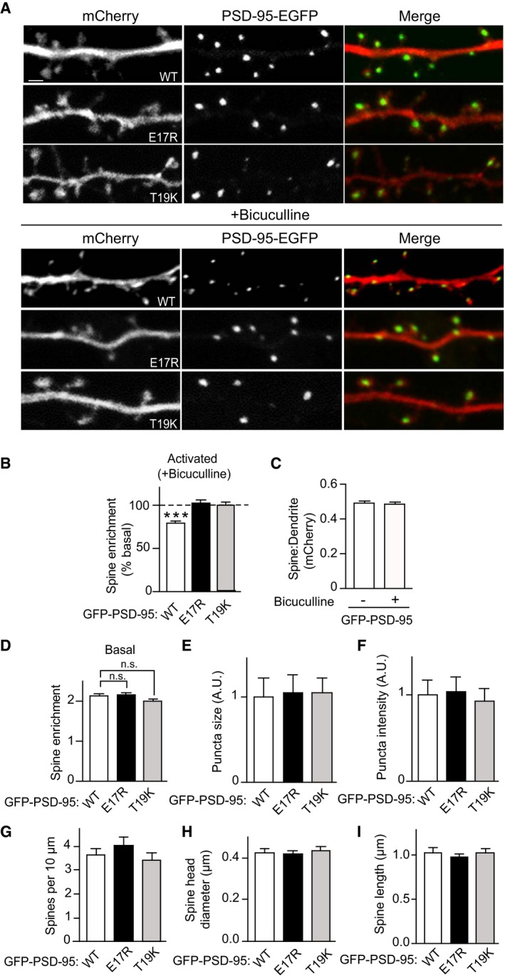
Cultured hippocampal neurons were co‐transfected with mCherry and EGFP‐tagged PSD‐95 wild type (WT), E17R, or T19K at 10–11 DIV and treated with BIC (50 μM) or left untreated at 17 DIV for 24–48 h before fixation.
-
ARepresentative confocal microscopic images of PSD‐95‐EGFP (green) and mCherry (red) used as “fill” to visualize the dendrite with spine heads (scale bar: 2 μm) after control (top) and BIC treatment (bottom). Individual channels are shown in grayscale.
-
BBIC treatment reduced spine enrichment of WT but not E17R or T19K PSD‐95 as quantified as the ratio of EGFP intensity in spine heads relative to that in the adjacent dendritic shaft. Values were normalized to control treatments, which were set to equal 100% for each PSD‐95 construct.
-
CmCherry showed no enrichment in spines and BIC had no effect on its localization.
-
D–FSpine enrichment and puncta size and intensity of PSD‐95 were comparable under basal conditions for WT, E17R, and T19K PSD‐95 (A.U., arbitrary unit).
-
G–ISpine density, head diameter, and length were comparable under basal conditions for neurons expressing WT, E17R, and T19K PSD‐95.