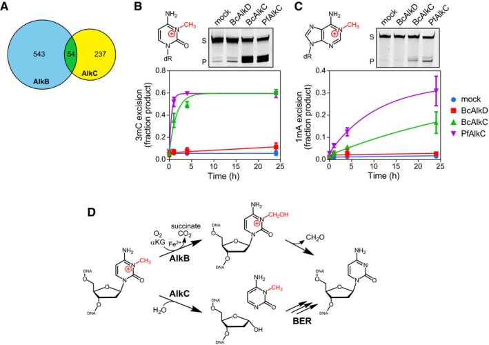
Figure 6. AlkC excises 3‐methylcytosine (3mC) and 1‐methyladenine (1mA) from DNA .

-
AVenn diagram of the numbers of bacterial species containing either AlkB (blue), AlkC (yellow), or both (green).
-
B, CChemical structures and in vitro base excision of 3mC (B) and 1mA (C) from 25‐mer double‐stranded oligodeoxyribonucleotides. Representative denaturing electrophoresis gels show substrate (S) and product (P) after a 24‐h incubation with either no enzyme (mock), BcAlkD, BcAlkC (AlkCα), or PfAlkC (AlkCβ). Plots show quantified time courses from three experiments (values are mean ± SD).
-
D3mC may be repaired in bacteria by either AlkB‐catalyzed oxidative demethylation or AlkC‐catalyzed base excision. αKG, α‐ketoglutarate.
