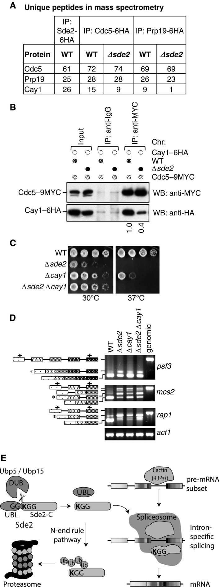
Figure 8. Sde2‐C facilitates association of Cay1 with spliceosomes.

- Sde2 Co‐IPs with Cactin in the spliceosome. Cdc5–6HA and Prp19‐6HA complexes were immunoprecipitated using anti‐HA antibody beads from Schizosaccharomyces pombe lysates wild‐type and Δsde2 strains. Co‐IP proteins were analysed by mass spectrometry. The table shows number of unique peptides obtained for each protein in mass spectrometry.
- Sde2 facilitates recruitment of Cay1 to the spliceosome. Assay is similar to (A). Lysates from S. pombe wild‐type and Δsde2 strain with Cdc5–9MYC and Cay1–6HA tagged were immunoprecipitated using anti‐MYC antibody beads. Co‐IP of Cay1–6HA was analysed by anti‐HA Western blot. Numbers below anti‐HA blot indicate ratio of Cay1 to Cdc5 (HA/MYC) signals obtained from ImageJ quantification of immunoblot signals.
- Sde2 and Cay1 are epistatic. Fivefold serial dilution spotting of indicated mutant strains was done on rich media. Plates were incubated at 30°C and 37°C until growth appeared.
- Cay1 and Sde2 knockout strains have similar pre‐mRNA splicing defects. Assay is similar as in Fig 5B.
- Schematics for Sde2 function and regulation.
Source data are available online for this figure.
