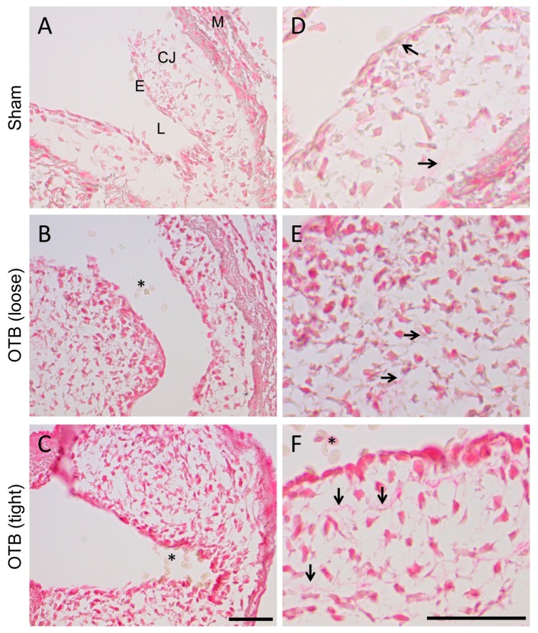
Figure 4.
Picrosirius red staining for collagen suggests increased collagen deposition in the banded OFT. (A–C) Photomicrographs of the OFT wall in sham and banded embryos. Collagen containing tissue is stained in red, and appears to increase with progressive band tightness. M, myocardium; CJ, cardiac jelly; E, endocardium; L, lumen. Scale, 50 μm. (D–F) Photomicrographs of the cardiac jelly suggest increased collagen fibrils (arrows) in the tightly banded OFT. Red blood cells (*) in the lumen are the negative control. Scale, 50 μm.

