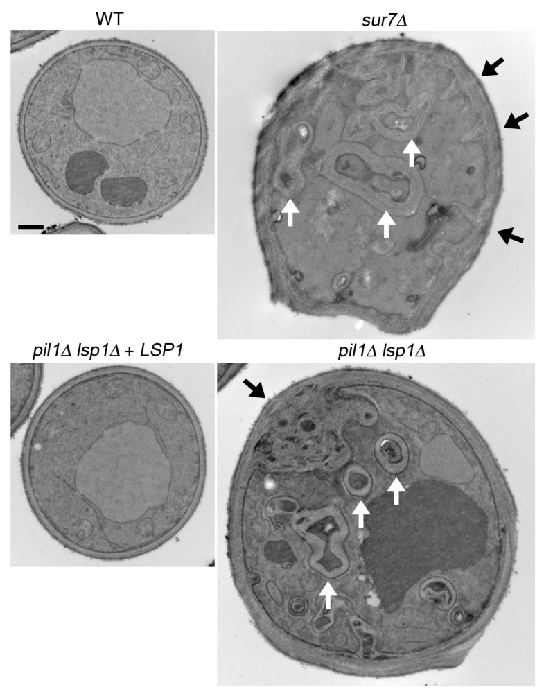
Figure 3.
Abnormal cell wall invaginations in C. albicans sur7∆ and pil1∆ lsp1∆ mutants. The indicated cell sections were analyzed by transmission electron microscopy. Thicker cell walls and invaginations of cell wall material were detected in both the sur7∆ mutant and the pil1∆ lsp1∆ double mutant. The white arrows indicate tubes of cell wall material. Black arrows indicate spots where there are spiky invaginations in the sur7∆ mutant and the large round cell wall invagination in the pil1∆ lsp1∆ mutant. Black bar, 1 µm. This image was reproduced from Figure 2 of the paper by Wang et al. [19].

